



Mapping Language And Memory Across The Brain With fMRI
Lateralization of Language and Memory: fMRI Contributions remains a valuable theme in cognitive neuroscience because it connects brain imaging with real-world assessment. Functional magnetic resonance imaging (fMRI) allows researchers to observe regional activity while people name objects, understand speech, encode information, or retrieve past experiences.
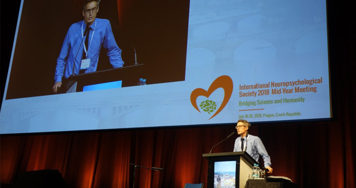
The subject also reflects the spirit of the INS 2018 meeting in Prague, held from July 18–20. The program brought together scientific advances, clinical neuropsychology, cultural awareness, and humane patient care—areas that are especially important when interpreting individual differences in brain organization.
Rather than treating language and memory as isolated abilities, modern neuropsychology examines their shared networks, timing, and adaptability. fMRI findings can guide assessment after neurological injury, support rehabilitation planning, and improve understanding of how the brain compensates when typical pathways are disrupted.
Why Hemispheric Specialization Matters
Lateralization describes the unequal contribution of the brain’s two hemispheres to a cognitive function. Language is often associated with the left hemisphere, particularly regions involved in speech production and comprehension. Memory, however, shows a more distributed pattern, with the medial temporal lobes, prefrontal cortex, and parietal regions contributing according to the type of task.
These patterns are tendencies rather than fixed rules. Age, handedness, developmental history, education, bilingualism, and neurological disease can all influence functional organization. A careful interpretation therefore considers the whole activation network instead of assigning a single ability to one brain region.
What Functional Imaging Reveals
During an fMRI experiment, changes in blood oxygenation provide an indirect measure of neural activity. Participants may read words, generate verbs, listen to sentences, encode pictures, or recognize previously presented material while the scanner records blood-oxygen-level-dependent signals.
Researchers compare task-related activity with a baseline condition and examine both localized responses and communication between regions. Functional connectivity analyses can reveal whether language areas work in concert with attention, executive-control, or memory systems. This network perspective is particularly useful when activation appears in unexpected locations.
Language Networks And Memory Systems
Classic language research highlights Broca’s area in the inferior frontal gyrus and Wernicke-related regions in the temporal cortex, yet contemporary models emphasize flexible, interacting circuits. Speech production, semantic processing, reading, and verbal working memory recruit partially overlapping pathways that extend into motor, auditory, and attentional networks.
Memory also depends on task demands. The hippocampus is central to forming and retrieving episodic memories, while frontal regions help organize strategies and monitor recall. Verbal encoding may engage left-sided networks, whereas visual-spatial memory often places greater demands on right-sided regions. These distinctions help explain why a person can show preserved learning in one modality and impairment in another.
| Cognitive process | Commonly observed emphasis | Important interpretation |
|---|---|---|
| Speech production | Left inferior frontal and motor regions | Activation varies with fluency, effort, and task design |
| Language comprehension | Temporal and frontal language networks | Meaning, syntax, and auditory demands recruit different areas |
| Verbal encoding | Left frontal and medial temporal regions | Strategy and familiarity can change the pattern |
| Episodic retrieval | Hippocampal and prefrontal systems | Successful recall depends on monitoring and context |
| Visual-spatial memory | Often greater right-hemisphere involvement | Performance also draws on attention and perception |
Individual Variation In Clinical Assessment
A strong fMRI study distinguishes normal variation from compensatory recruitment. After stroke, traumatic brain injury, epilepsy, or tumor treatment, language functions may shift toward homologous regions in the opposite hemisphere or become more distributed across both sides. Increased activation does not automatically mean better performance; it may represent additional effort.
Clinical teams combine imaging with behavioral measures, interviews, and standardized neuropsychological tests. This multimethod approach avoids overinterpreting a colorful activation map and helps connect brain activity with communication, learning, daily functioning, and quality of life. For patients recovering from injury, research on veteran brain rehabilitation illustrates why functional outcomes must remain central.
Cultural And Methodological Considerations
Language-related activation is shaped by the language a person speaks, literacy experience, cultural context, and familiarity with testing materials. Bilingual participants may use overlapping or partly distinct neural systems depending on proficiency, age of acquisition, and the situation in which each language was learned.
Experimental design also matters. Small samples, motion artifacts, multiple comparisons, scanner differences, and poorly matched control tasks can weaken conclusions. Researchers should report participant characteristics clearly and interpret hemispheric asymmetry alongside performance accuracy, response time, and subjective difficulty.
Applying Findings To Practice
fMRI is most useful when it answers a precise clinical or research question. Practical applications include presurgical language mapping, investigation of memory disorders, monitoring recovery, and studying treatment-related neural plasticity. It can also clarify whether a patient’s difficulty reflects impaired storage, inefficient retrieval, limited attention, or language-processing barriers.
Useful principles for interpreting imaging evidence include:
- Pair activation maps with detailed cognitive and functional assessment.
- Treat hemispheric dominance as a measurable pattern, not a permanent label.
- Account for age, handedness, bilingualism, education, and cultural background.
- Examine connectivity and task performance alongside regional activation.
- Use repeated or multimethod evaluation when recovery or decline is changing over time.
The continuing value of this field lies in its ability to connect neuroscience with compassionate care. As imaging methods become more precise, their greatest contribution will come from helping clinicians understand each person’s strengths, limitations, and capacity for adaptation.
Explore the INS 2018 scientific materials and related neuropsychological resources to see how brain imaging can inform responsible assessment, rehabilitation, and patient-centered practice.
General Information
Important information about the meetingIndustry
Support and exhibition opportunitiesCzech Republic
Beautiful country situated in the very heart of EuropeContact
How can we help you?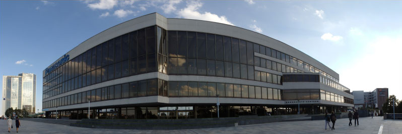
Prague Congress Centre (KCP)
5.května 65140 21 Prague 4
Czech Republic
Phone: +420 261 171 111
Website: www.kcp.cz