



Using fMRI to Predict Recovery After Brain Injury
Recovery after traumatic brain injury, stroke, or acquired brain injury can follow very different paths. Two people with similar lesions may show contrasting changes in memory, attention, language, movement, or emotional regulation. Functional magnetic resonance imaging (fMRI) offers one way to study this variation by showing how brain networks respond during tasks or at rest.
Rather than treating a scan as a forecast written in advance, clinicians and researchers use fMRI as one component of a broader neuropsychological picture. Its value lies in connecting brain activity with behavior, medical history, rehabilitation access, and the person’s everyday goals.
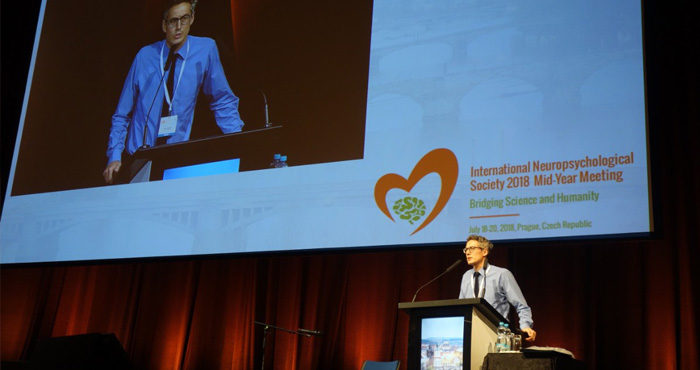
The scientific and humane themes associated with the INS 2018 meeting in Prague remain relevant: advanced neuroscience becomes most useful when it improves assessment, communication, and patient care. Predictive imaging should therefore support clinical judgment rather than replace it.
What fMRI Reveals After Injury
Traditional structural MRI shows damaged tissue, swelling, bleeding, or changes in anatomy. fMRI measures changes in blood oxygenation associated with neural activity. During a task-based scan, researchers may observe which regions are engaged while a person remembers words, moves a hand, or understands speech.
Resting-state fMRI provides a different perspective. It measures synchronized activity between brain regions when the person is not completing a specific task. These patterns can reveal changes in functional connectivity, including weakened communication within a network or increased cooperation between regions that were previously less involved.
Why Network Activity Matters
Brain injury often disrupts networks rather than one isolated location. Language, executive control, memory, and motor abilities depend on coordinated activity across several regions. A scan may therefore show that recovery involves the reorganization of existing pathways, recruitment of nearby areas, or support from the opposite hemisphere.
Researchers may combine fMRI with diffusion imaging, electroencephalography, lesion mapping, and repeated behavioral testing. This multimodal approach can distinguish temporary changes from durable adaptation. A single scan is less informative than a series of measurements linked to real improvements in daily functioning.
From Brain Signals To Recovery Estimates
Predictive models may use activation strength, connectivity, lesion location, age, initial impairment, and rehabilitation data to estimate likely outcomes. For example, preserved connectivity in a language network could be associated with better naming recovery, while intact motor pathways might help explain future gains in walking or upper-limb control.
These estimates are probabilities, not promises. Prediction is most useful when it identifies a realistic range of outcomes and helps clinicians plan therapy intensity, communication supports, and follow-up assessments. It should never be used to deny rehabilitation because a statistical model assigns a low probability of improvement.
| fMRI Approach | What It Can Show | Clinical Value | Important Limitation |
|---|---|---|---|
| Task-based fMRI | Activity during language, memory, or movement tasks | Identifies engaged or preserved systems | Requires reliable task performance |
| Resting-state fMRI | Connectivity among functional networks | Useful when testing is difficult | Results can vary with motion and analysis methods |
| Longitudinal fMRI | Changes across rehabilitation | Tracks reorganization over time | Requires repeated scans and consistent protocols |
| Combined imaging | Relationships between structure and function | Produces a fuller recovery profile | More expensive and technically demanding |
The Role Of Patient Context
Imaging findings must be interpreted alongside education, language, culture, mood, fatigue, medication, and premorbid ability. A task designed for one linguistic or cultural group may underestimate another person’s skills. Guidance on culturally responsive assessment is therefore directly relevant to any imaging-informed prediction.
The scan environment can also influence results. Pain, anxiety, unfamiliar instructions, hearing difficulties, and limited literacy may alter task performance or create excessive head movement. Clinicians should document these factors instead of treating every difference in activation as evidence of neurological dysfunction.
Limits Of Neuroimaging Prediction
fMRI has technical weaknesses, including limited temporal resolution, sensitivity to motion, variable preprocessing choices, and differences between scanners. People may also activate compensatory regions without achieving meaningful functional improvement. Conversely, a person may improve through strategies that are not clearly reflected in a conventional activation map.
Ethical issues are equally important. Predictive findings can affect decisions about work, education, insurance, or long-term care. Patients should receive clear explanations of uncertainty, data privacy, and the difference between research findings and validated clinical tools. Developments in related areas, such as the dementia diagnosis advances, illustrate why promising biomarkers still require careful validation before routine use.
Applying Findings In Rehabilitation
The strongest clinical pathway links imaging to an individual rehabilitation plan. A preserved network may suggest which abilities can be trained directly, while evidence of compensation may encourage strategy-based therapy. Repeated neuropsychological testing remains essential for determining whether those plans produce meaningful changes.
Useful practice principles include:
- Combine fMRI with structural imaging, behavioral testing, and functional reports.
- Treat predictions as changing estimates that should be updated over time.
- Account for language, culture, fatigue, mood, and access to rehabilitation.
- Explain uncertainty in plain language before using results in care planning.
- Measure outcomes in everyday activities, not only in the scanner.
When these safeguards are followed, fMRI can help connect neuroscience with compassionate clinical decision-making. The goal is not to label a person’s future, but to identify opportunities for recovery and tailor support around the abilities that matter most.
Explore the INS 2018 journal and conference resources to deepen understanding of neuropsychological assessment, brain-network science, and patient-centered care. Building responsible bridges between imaging research and rehabilitation can make recovery planning more precise while keeping the person—not the scan—at the center.
General Information
Important information about the meetingIndustry
Support and exhibition opportunitiesCzech Republic
Beautiful country situated in the very heart of EuropeContact
How can we help you?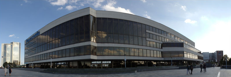
Prague Congress Centre (KCP)
5.května 65140 21 Prague 4
Czech Republic
Phone: +420 261 171 111
Website: www.kcp.cz